
Research Review: Changes in Cervical Spine Musculature Observed with Faulty Prone Hip Extension Pattern
By Nicholas Rolnick SPT, MS, CSCS
Edited by Brent Brookbush DPT, PT, COMT, MS, PES, CES, CSCS, ACSM H/FS
Original Citation: Bruno PA, Murphy DR. (2011). An investigation of neck muscle activity in asymptomatic participants who show different lumbar spine motion patterns during prone hip extension. Journal of Manipulative and Physiological Therapeutics. 34(8): 525-532. - Abstract
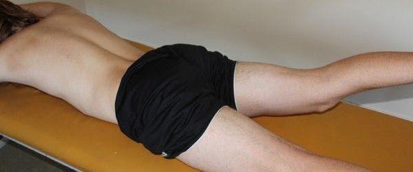
The prone hip extension pattern tested during the current study's protocol - http://www.hanslindgren.com/blog/janda-s-pelvic-cross-syndrome/
Why is this relevant?: The classic prone hip extension pattern test (PHE) used to assess neuromuscular coordination of the lumbopelvic complex lacks clinical validity in research trials, raising questions as to its usefulness in practice. In the PHE, the examiner places his or her hands on the same-side hamstring , gluteus maximus , and erector spinae musculature and asks the patient/client to extend the same-side leg into the air. The examiner assesses for relative activation patterns in the palpated musculature for insight into muscular recruitment order. It has been hypothesized that there exists abnormal recruitment and delayed gluteus maximus activation (hamstrings firing before gluteus maximus , erector spinae firing before gluteus maximus ) in individuals with low back pain (1), but specific criteria defining what is "normal" and "abnormal" have yet to be definitively established. The authors of the current study sought to increase clinical validity of the PHE by providing a criteria for what is "normal" and "abnormal." Further, research has suggested the presence of altered cervicothoracic muscle activation patterns (cervical erector spinae , upper trapezius , levator scapulae , rhomboids ) in individuals with neck pain (2) during the PHE. The current study investigated differences in activity patterns between cervical erector spinae and upper trapezius musculature in asymptomatic "normal" and "abnormal" individuals without neck pain during a prone hip extension exercise.
Study Summary
| Study Design | Observational |
| Level of Evidence | Level 2 |
| Subject Demographics |
|
| Outcome Measures | |
| Results |
Mean Normalized Muscle Activities for the Ipsi ES, Contra ES, Ipsi UT, and Contra UT in "positive" and "negative" groups
|
| Conclusions | The presence of one or more of 3 abnormal lumbar spine movements in the PHE test is associated with an increase in the activity of the ipsilateral upper trapezius on the side of the hip extension in asymptomatic participants. |
| Conclusions of the Researchers | The results of the current study seem to suggest that abnormal lumbar spine movements results in compensatory muscle activation pattern that includes the upper trapezius. |
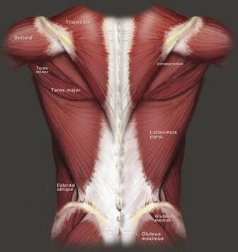
Note the muscular and fascial attachments to the spine; Imagine how abnormal movements of the spine would cause other muscles to reflexively contract to stabilize- http://dragongatefitness.files.wordpress.com/2012/11/backanat.jpg
Review & Commentary:
The current study methodology was simple and adequately designed to test the hypothesis that abnormal movements of the lumbar spine would elicit changes in upper cervical spine muscle activity. The methods were detailed and well described for replication in future studies on the topic. The exclusion criteria was comprehensive to ensure that the participant pool in the study were truly "asymptomatic." The foundation of the study relied upon establishment of a new criteria to evaluate the PHE test, independent of palpation of the hip and back muscular. The authors described three faulty movement patterns associated with the lumbar spine in the PHE, had a qualified clinician (Chiropractor) evaluate every participant, and performed blinding on the data analysis, minimizing bias. The authors of the current study plan to utilize the experimental protocol for a future study on individuals with neck pain to provide data for comparison.
The current study has limitations that should be considered before application to practice. First, the PHE pattern was originally popularized in the literature due to its hypothesized relationship to gait, that is, similar recruitment of the muscles of the hip would be used during the PHE and active hip extension during gait. However, the literature is conflicting on this topic, questioning the correlation between the PHE and gait, and further muscle recruitment during functional activities. Second, although the authors created a new system for classification of abnormal lumbar spine motion, quantification of the amount of motion and its relation pain was not established (participants in this study were asymptomatic). Further, it is not known whether techniques aimed at restoring neuromuscular coordination of the lumbopelvic complex will affect test results in individuals exhibiting abnormal motion, and whether those results will be representative of changes in functional tasks.
Why is this study important?
The current study is evidence that abnormal motion of the spine may influence distal muscle activation patterns. This study investigated how the presence of one or more of three described faulty lumbar extension movement patterns influenced muscle activation of the cervical erector spinae and upper trapezius musculature. Increased upper trapezius activity was observed on the same side as the faulty hip extension pattern. Other research has shown changes in muscle activity of the cervical spine in individuals with chronic neck pain (3). Further research is needed to clarify the relationship between hip, lumbar spine and cervical dysfunction.
How does it affect practice?
The importance of optimal motion of the spine, its influence on proximal and distal muscular activity, and how it is correlated with pain and performance has yet to be fully elucidated in the literature. The current study adds to a growing body of evidence that abnormal motion of the spine may have an affect on muscle activity distal to the site of dysfunction (excessive ipsilateral upper trapezius activation, in this study); a phenomenon sometimes referred to as "regional interdependence". Whether or not regional interdependence contributes to onset of pain has yet to be investigated, but dysfunctional movement has been linked to pain in several studies (4-12).
Relative to this study, dysfunction related to the hip and/or lumbar spine may result in dysfunction of the cervical spine, which may warrant assessment and correction of all segments exhibiting dysfunction. In a previous research review, it was found that correcting forward head posture in lumbar spine patients had a positive affect on long-term outcomes - Moustafa et al.
How does it relate to Brookbush Institute Content?
In all predictive models of postural dysfunction proposed by the Brookbush Institute (lumbopelvic hip complex dysfunction (LPHCD) sacroiliac joint dysfunction (SIJD) , lower leg dysfunction (LLD) , and upper body dysfunction (UBD) ), regional interdependence is assumed. Further, changes in the activity of core sub-systems , altered myofascial activity and length, arthrokinematic dyskineisis, altered force transmission and the potential for pain and risk of injury is discussed. In the current study, excessive and/or suboptimal movement of the lumbopelvic hip complex in a prone hip extension exercise resulted in increased ipsilateral cervical muscle activity. It can be postulated that the increase in upper trapezius activation is compensatory, along with over-activity of the deep longitudinal and anterior oblique subsytem , for a relatively under-active intrinsic stabilization subsystem (ISS) and posterior oblique subsystem (POS) .
With respect to the core subsystems, the Brookbush Institute recommends core activation and subsystem integration exercises to address altered activity and recruitment, following mobility techniques, and activation techniques.
For example, following mobility techniques, to increase the activity of the ISS , transverse abdominis activation may be used. This may be followed by gluteus maximus activation , a bridge progression for integration of core musculature, following by an exercises that include lower body and an upper body pull component to integrate the POS .
The following videos are basic steps to activating and integrating the commonly underactive ISS and POS , improving lumbopelvic spine motion and stability during functional exercise.
Brookbush Institute Videos
Review of Core Subsystems
Transverse Abdominis Isolated Activation - Intrinsic Stabilization Subsystem
Transverse Abdominis and Gluteus Maximus Activation - Intrinsic Stabilization Subsystem Progressions
Ball Bridge - (Progression off bridging exercise) - Posterior Oblique Subsystem
Squat to Row - Posterior Oblique Subsystem Integration
Bibliography:
- Hungerford B, Gilleard W, Hodges P. (2003). Evidence of altered lumbopelvic muscle recruitment in the presence of sacroiliac joint pain. Spine, 28 (14): 1593–1600.
- Murphy D. 2000. Evaluation of posture and movement patterns related to the cervical spine. In: Murphy D, editor. Conservative management of cervical spine syndromes. New York: McGraw-Hill, 307-327.
- Jull GA. (2000). Deep cervical flexor muscle dysfunction in whiplash. Journal of Musc Pain. 8(1/2): 143-154.
- Helgadottir, H., Kristjansson, E., Einarsson, E., Karduna, A., & Jonsson, H. (2011). Altered activity of the serratus anterior during unilateral arm elevation in patients with cervical disorders. Journal of electromyography and kinesiology,21(6), 947-953.
- Ayhan, C., Camci, E., & Baltaci, G. (2015). Distal radius fractures result in alterations in scapular kinematics: A three-dimensional motion analysis. Clinical Biomechanics.
- José Miota Ibarra, Hong-You Ge, Chao Wang, Vicente Martínez Vizcaíno, Thomas Graven-Nielsen, Lars Arendt-Nielsen – The Journal of Pain, Volume 12, Issue 12, December 2011, Pages 1282–1288
- Apkarian, A.V., Sosa, Y. Sonty, S., Levy, R.M., Harden, R.N., Parrish, R.B., and Gitelman, D.R. (2004) Chronic back pain is associated with decreased prefrontal and thalamic gray matter density. The Journal of Neuroscience. 24 (46): 10410-10415
- Hodges, P., Richardson, C. (1996). Inefficient Muscular Stabilization of the Lumbar Spine Associated With Low Back Pain: A Motor Control Evaluation of Transverse Abdominis. Spine, 21(22), 2640-2650.
- Thigpen CA, Padua DA, Michener LA, Guskiewicz K, Giuliani C, Keener JD, Stergiou N. (2010). Head and shoulder posture affect scapular mechanics and muscle activity in overhead tasks. Journal of Electromyography and Kinesiology. 20: 701-709.
- Ireland, ML., Wilson, JD., Ballantyne, BT., Davis, IM. (2003). Hip Strength in Females With and Without Patellofemoral Pain. J Orthop Sports Phys Ther 2003. 33: 671-676.
- Franettovich, S. M., Honeywill, C. O. N. O. R., Wyndow, N., Crossley, K. M., & Creaby, M. W. (2014). Neuromotor control of gluteal muscles in runners with achilles tendinopathy. Medicine and science in sports and exercise, 46(3), 594-599.
- Hungerford, B., Gilleard, W., Hodges, P. (2003) Evidence of altered lumbopelvic muscle recruitment in the presence of sacroiliac joint pain. Spine 28(14), 1593-1600
- Moustafa, I. M., & Diab, A. A. (2015). The Effect of Adding Forward Head Posture Corrective Exercises in the Management of Lumbosacral Radiculopathy: A Randomized Controlled Study. Journal of manipulative and physiological therapeutics, 38(3), 167-178
© 2016 Brent Brookbush
Questions, comments, and criticisms are welcomed and encouraged -