
Research Review: Electromyographic Analysis of Hip Rehabilitation Exercises in a Group of Healthy Subjects
By Tristan Rodik, M.AT., ATC
Edited By Brent Brookbrush, DPT, PT, COMT, MS, PES, CES, CSCS, ACSM H/FS
Original Citation: Bolgla, L. A. and Uhl, T. L. (2005) Electromyographic analysis of hip rehabilitation exercises in a group of healthy subjects. Journal of Orthopaedic and Sports Physical Therapy, 35(8), 487-494. ABSTRACT
Why the Study is Relevant: Hip abductor weakness is correlated with low-back pain, patellofemoral pain syndrome, Achilles tendinopathy, and optimal function of the lower extremity and lumbopelvic hip complex (1-10). Human movement professionals in rehab and performance settings use a wide variety of exercises to address hip abductor weakness. This 2005 study by Bolgla et al. adds to a growing body of research using electromyography to compare gluteus medius strengthening exercises (11-14). Findings suggest that weight-bearing exercises result in a greater percentage of maximum voluntary isometric contraction (%MVIC) than non-weight-bearing exercises. "Pelvic drops" produced the greatest %MVIC, and side-lying abduction produced the greatest %MVIC of the non-weight bearing exercises. The findings may aid the human movement professional in selecting and progressing exercise.
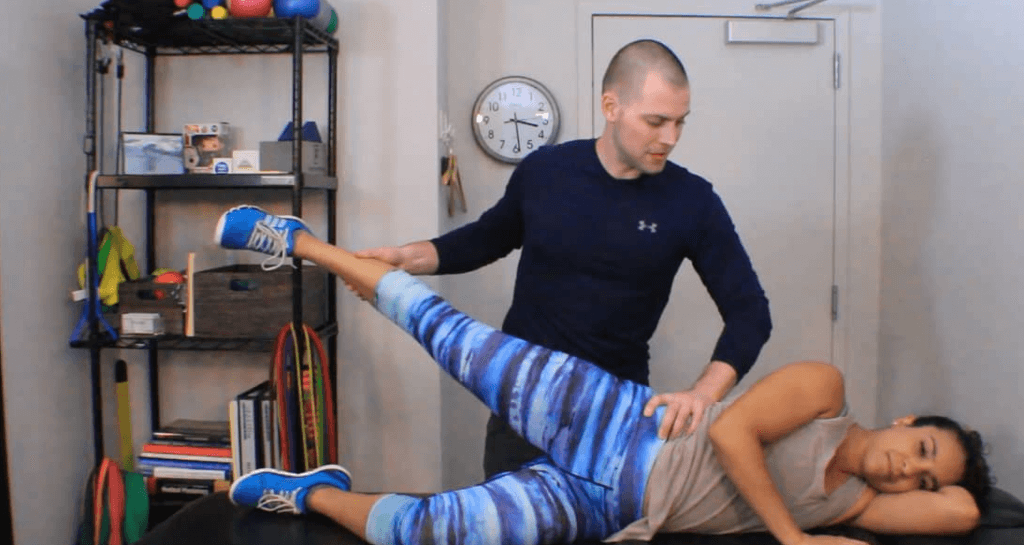
Gluteus Medius Manual Muscle Testing
| Study Design | Single-occasion, repeated-measures design |
| Level of Evidence | III Evidence from non-experimental descriptive studies |
| Subject Characteristics | Demographics
Inclusion Criteria:
Exclusion Criteria:
|
| Methodology | Prior to Testing:
Exercise Testing Procedure:
The Tested Exercises:
|
| Data Collection and Analysis |
|
| Outcome Measures |
|
| Results | The six selected exercises are listed below from highest to lowest %MVIC for the right gluteus medius:
|
| Our Conclusions | This study measured the levels of demand that various hip abductor exercises place on the gluteus medius The findings provide human movement professionals with guidance on how to safely and effectively progress a client through a gluteus medius strengthening program based on initial strength and health status. |
| Researchers' Conclusions
|
Less EMG activity was noted among the NWB exercises when compared to the WB exercises. The NWB exercises may be considered for clients who cannot safely perform the WB exercises. Since the WB exercises are more demanding and challenge the gluteus medius's ability to stabilize the pelvis in WB exercises (its primary functional role), programs should progress towards WB exercises for all capable clients. The findings can be used in designing an exercise protocol and exercise progression for hip abductor strengthening. |
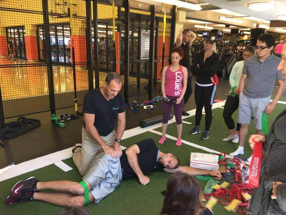
Karl Sterling, BI Presenter prepping to teach Gluteus Medius Activation
Review and Commentary:
This study adds to the growing body of research comparing gluteus medius strengthening exercises (11-14). Greater percentage of maximal voluntary isometric contraction (%MVIC) was recorded during weight-bearing (WB) exercises, which may be due to the increased external torque generated by the body's mass and additional stability demands. Side-lying abduction produced the greatest %MVIC of the non-WB exercises, which also creates the largest moment arm and external torque of the non-WB exercises. These findings provide human movement professionals with guidance to determine optimal exercise selection and progression.
This study had many methodological strengths, including:
- The results may provide a progression of the six exercises relative to gluteus medius %MVIC.
- The use of EMG provides human movement professionals with quantifiable evidence to support recommendations for hip abduction strengthening exercises.
- The WB exercises may have transference to the day-to-day hip stabilization requirements for activities of daily living.
- Reliability among trials was evaluated to assess consistency among participants by evaluating the intra-class correlation coefficient (ICC). The ICC was 0.92, inferring minimal electrode movement and consistent effort with each exercise.
Weakness that should be noted prior to clinical integration:
- Only healthy participants were included. It is unclear whether the gluteus medius will exhibit similar activation patterns in injured populations.
- During the WB exercises, participants were instructed to maintain a level pelvic position. However, this was not objectively monitored by the researchers, which may have led to varied postures (and potentially altered muscular activity).
- Only the right extremity was tested and leg dominance was not noted. It is unclear if this may have influenced the findings.
- The gluteus medius was the only hip abductor muscle evaluated. Future research should evaluate all hip abductor muscles - including the gluteus maximus , gluteus minimus , and tensor fascia latae - during hip abduction exercises (15).
Why This Study is Important:
The importance of gluteus medius strengthening and its correlation to lower-extremity function and pain (such as femoral adduction and knee valgus) has been well established (1-10). This study evaluated gluteus medius activity in six commonly used hip abductor strengthening exercises, including non-weight bearing and weight bearing variations. The findings provide guidance for human movement professionals when determining exercise progression for gluteus medius strengthening. Additional note: more research should be done to compare techniques with similar intent in pursuit of determining optimal exercise selection and practice.
How the Findings Apply to Practice:
The exercises examined in this study are commonly used by human movement professionals and provide valuable insight on %MVIC (and potentially torque demand differences) between weight-bearing (WB) and non-weight-bearing (NWB) exercises. While the NWB exercises involve controlling the limb and added resistance, the WB exercises must control the head, arms, trunk and NWB leg increasing external load and stability demand. This information may be considered by human movement professionals when designing an exercise intervention as well as exercise progression.
Progression of Exercise based on %MVIC (Least to Highest Activity):
- NWB standing flexed-hip hip-abduction: 28 +/- 21
- NWB standing hip-abduction: 33 +/- 23
- NWB Side-lying hip-abduction: 42 +/- 23
- WB hip-abduction: 42 +/- 27
- WB standing hip-abduction with flexed hip: 46 +/- 34
- Pelvic Drop: 57 +/- 32
How this Study Relates to Brookbush Institute Content:
The Brookbush Institute (BI) recognizes the importance of strong hip abductors, specifically the gluteus medius for optimal movement. This muscle is commonly classified as long/under-active in the predictive models of postural dysfunction - Lower Extremity Dysfunction (LED) , Lumbo-Pelvic-Hip Complex Dysfunction (LPHCD) , and Sacroiliac Joint Dysfunction (SIJD) . This study supports the use of both non-weight bearing (NWB) and weight-bearing (WB) exercises for the gluteus medius , perhaps as a progression from NWB exercises to WB exercises. The findings of this study are similar to the BI's corrective exercise template and activation circuits for the gluteus medius . The subtle difference in the BI model includes consideration of "reactive activation" to improve firing rate and sequence, and "subsystem integration " to improve whole-body movement patterns.
Functional Anatomy of the Gluteal Muscle Group:
Manual Muscle Testing of the Hip Abductors:
Isolated Activation for the Gluteus Medius:
Side-lying Gluteus Medius Exercises:
Gluteus Medius Reactive Integration Progressions:
Gluteus Medius Reactive Integration Side-stepping Progressions:
Gluteus Medius Reactive Integration Side-stepping (Part II) Progressions:
Gluteus Medius Reactive Integration Side-hoping Progressions:
Recommended Readings:
- Manual Muscle Testing for an Active Population: Lower Body
- Introduction to the Overhead Squat Assessment
- Overhead Squat Assessment Solutions Table - Sign Clusters and Compensation Patterns :
- Gluteus Medius Activation
Bibliography:
- Dos Reis, A. C., Ferrari Correa, J. C., Bley, A. S., Rabelo, N. D., Fukuda, T. Y., and Lucareli, P. R. (2015) Kinematic and kinetic analysis of the single-leg triple hop test in women with and without patellofemoral pain. Journal of Orthopaedic and Sports Physical Therapy, 45(10), 799-807
- Noehren, B., Hamill, J., and Davis, I. (2013) Prospective evidence for a hip etiology in patellofemoral pain. Medicine and Science in Sports and Exercise, 45(6), 1120-1124
- Ireland, M. L., Willson, J. D., Ballantyne, B. T., and Davis, I. M. (2003) Hip strength in females with and without patellofemoral pain. Journal of Orthopaedics and Sports Physical Therapy, 33(11), 671-676
- Noehren, B., Scholz, J., and Davis, I. (2011) The effect of real-time gait retraining on hip kinematics, pain and function in subjects with patellofemoral pain syndrome. British Journal of Sports Medicine, 45, 691-696
- Franettovich Smith, M. M., Honeywill, C., Wyndow, N., Crossley, K. M., and Creaby, M. W. (2014) Neuromotor control of gluteal muscles in runners with achilles tendinopathy. Medicine and Science in Sports and Exercise, 46(3), 594-599
- Souza, R. B. and Powers, C. M. (2009) An evaluation of hip strength and femoral structure in women with and without patellofemoral pain. The American Journal of Sports Medicine, 37(3), 579-587
- Ramskov, D., Barton, C., Nielson, R. O., and Rasmussen, S. (2015) High eccentric hip abduction strength reduces the risk of developing patellofemoral pain among novice runners initiating a self-structured running program: a 1-year observational study. Journal of Orthopaedic and Sports Physical Therapy, 45(3), 153-161
- Boling, M. C., Padua, D. A., and Creighton, R. A. (2009) Concentric and eccentric torque of the hip musculature in individuals with and without patellofemoral pain. Journal of Athletic Training, 44(1), 7-13
- Cooper, N. A., Scavo, K. M., Strickland, K. J., Tipayamongkol, N., Nicholson, J. D., Bewyer, D. C., and Sluka, K. A. (2016) Prevalence of gluteus medius weakness in people with chronic low back pain compared to healthy controls. European Spine Journal, 25(4), 1258-1265
- Smith, J. A., Popovich, J. M. and Kulig, K. (2014) The influence of hip strength on lower-limb, pelvis, and trunk kinematics and coordination patterns during walking and hopping in healthy women. Journal of Orthopaedics and Sports Physical Therapy, 44(7), 525-531
- Boren, K., Conrey, C., Le Coguic, J., Paprocki, L., Voight, M., and Kevin Robinson, T. (2011) Electromyographic analysis of gluteus medius and gluteus maximus during rehabilitation exercises. International Journal of Sports Physical Therapy, 6(3), 206-223
- McBeth, J. M., Earl-Boehm, J. E., Cobb, S. C., and Huddleston, W. E. (2012) Hip muscle activity during 3 side-lying hip-strengthening exercises in distance runners. Journal of Athletic Training, 47(1), 15-23
- Berry, J. W., Lee, T. S., Foley, H. D., and Lewis, C. L. (2015) Resisted side stepping: the effect of posture on hip abductor muscle activation. Journal of Orthopaedic and Sports Physical Therapy, 45(9), 675-682
- Distefano, L. J., Blackburn, J. T., Marshall, S. W., and Padua, D. A. (2009) Gluteal muscle activation during common therapeutic exercises. Journal of Orthopaedic and Sports Physical Therapy, 39(7), 532-540
- Flack, N. A., Nicholson, H. D., and Woodley, S. J. (2012). A review of the anatomy of the hip abductor muscles, gluteus medius, gluteus minimus, and tensor fascia lata. Clinical Anatomy, 25(6), 697-708
© 2017 Brent Brookbush
Questions, comments, and criticisms are welcomed and encouraged.