
Research Review: Roentgenographic findings in the cervical spine in asymptomatic persons: A ten-year follow-up.
By Amy Martinez DPT, PT
Edited by David Boettcher MSc, BA, NASM CPT, PES & CES & NPTI
Edited by Brent Brookbush DPT, PT, COMT, MS, PES, CES, CSCS, ACSM H/FS
Original Citation:
Gore, R. (2001). Roentgenographic findings in the cervical spine in asymptomatic persons: A ten-year follow-up. Spine, 26 (22), 2463-2466. -ABSTRACT
Introduction:
Research suggests that 20% of the population may experience neck pain at some point in their lives (1-4). Research has demonstrated correlation between neck pain and cervical malalignment (5), changes in scapular mechanics and alterations in activity of the trapezius and serratus anterior (8,9). This 2001 prospective study, by an American researcher, established a correlation between chronic neck pain, cervical spine subluxations and degenerative changes. The findings suggest cervical joint mobility should be assessed in those who exhibit signs of neck pain, cervical dysfunction and/or upper body dysfunction (UBD) .
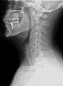
Lateral view of cervical spine x-ray - By Stillwaterising - Own medical image, work for hire, CC0 1.0 Universal Public Domain Dedication, https://commons.wikimedia.org/wiki/File:Cervical_Xray_Lateral_View.jpg
Study Summary
| Study Design | Prospective Study |
| Level of Evidence | III - Evidence from non-experimental descriptive studies, such as comparative studies, correlation studies, and case-control studies |
| Participant Characteristics | Demographics
Inclusion Criteria:
Exclusion Criteria:
|
| Methodology |
|
| Data Collection and Analysis | Data Collection
Data Analysis
|
| Outcome Measures |
|
| Results | Degeneration:
Pain
|
| Researchers' Conclusions | The number of subluxations and severity of degenerative changes in the cervical spine increased with age. Over a 10-year period, 15% of the participants reported developing pain, which correlates with additional research (1-4). Degenerative changes at C6-C7 visible on an initial X-ray was a statistically significant predictor of pain. |

Cervical Distraction Test
How this study contributes to the body of research:
This prospective study compared the lateral x-rays of 159 asymptomatic participants with 10-year follow-up. Outcomes included degrees of cervical lordosis, degenerative changes and incidence of self-reported neck pain. A previous cross-sectional study demonstrated a correlation between the level of spinal degeneration and chronic neck pain (10). Unique to this study, participants were initially asymptomatic and re-evaluated ten years later, allowing for correlations to be determined between the assessed signs of degeneration and increased risk of future neck pain.
How the Findings Apply to Practice:
The findings of this prospective study imply a correlation between cervical degenerative changes and increased incidence of neck pain, especially when degenerative changes are noted at the C6-C7 level. This may suggest that it is possible to develop screening methods that would be predictive of future pain and injury, based on measurable impairments (e.g. range of motion) correlated with these dysfunctions. Human movement professionals should consider the addition of cervical movement assessments and techniques for addressing cervical impairments prior to the onset of pain and injury.
Strengths
- Prospective studies are relatively rare in human movement science. This study began with asymptomatic individuals and followed up ten-years later, providing evidence that impairment may be a cause and/or a predictor of pain.
- The researcher investigated correlations between findings and age, to aid in isolating predictive factors from age-related changes.
- The researcher was blind to the participant's pain status at the time time of the X-ray, minimizing tester bias.
Weakness and limitations
- At the follow-up session participants were asked if they had experienced pain over the ten-year period. This long-time period may have introduced errors due to memory recall. A more frequent follow-up, or series of follow-ups, may help to improve the results.
- The researcher evaluated lateral X-rays, which limits evaluation of facet changes; additional X-ray angles and functional evaluations may enhance predictive reliability and clinical applicability.
- The term "subluxation" is not well defined in this study.
How the study relates to Brookbush Institute Content?
The Brookbush Institute (BI) continues to refine and develop predictive models of dysfunction, including Cervicothoracic Dysfunction (CTD) and Upper Body Dysfunction (UBD) , with the intent of optimizing evidence-based education and practice. This study supports the assertion that dysfunction is predictive and perhaps causative of cervical spine pain. The BI will continue to pursue optimal practice by integrating research, with practical application and outcomes, in pursuit of refining practice.
The following videos illustrate some techniques commonly recommended by the Brookbush Institute for Upper Body Dysfunction and Deep Cervical Flexor Activation:
Cervical Lateral Flexion Goniometry
Cervical Distraction Test
Cervical Fascia Instrument Assisted Soft-Tissue Manipulation
Cervical Spine Posterior to Anterior Mobilization
Cervical Manipulation
Deep Cervical Flexor Isolated Activation
Combined Deep Cervical Flexor and External Rotator Activation
Deep Cervical Flexor Reactive Activation
Bibliography:
- Bovim, G., Schrader, H., & Sand, T. (1994). Neck pain in the general population. Spine, 19(12), 1307-1309.
- Côté, P., Cassidy, J. D., Carroll, L. J., & Kristman, V. (2004). The annual incidence and course of neck pain in the general population: a population-based cohort study. Pain, 112(3), 267-273.
- Mäkela, M., Heliövaara, M., Sievers, K., Impivaara, O., Knekt, P., & Aromaa, A. (1991). Prevalence, determinants, and consequences of chronic neck pain in Finland. American journal of epidemiology, 134(11), 1356-1367.
- Picavet, H. S. J., & Schouten, J. S. A. G. (2003). Musculoskeletal pain in the Netherlands: prevalences, consequences and risk groups, the DMC3-study. Pain, 102(1-2), 167-178.
- Bogduk, N. (1995). The anatomical basis for spinal pain syndromes. Journal of Manipulative and Physiological Therapeutics, 18(9), 603-605.
- Falla, D., Jull, G., & Hodges, P. W. (2004). Feedforward activity of the cervical flexor muscles during voluntary arm movements is delayed in chronic neck pain. Experimental brain research, 157(1), 43-48.
- Falla, D., O’Leary, S., Farina, D., & Jull, G. (2011). Association between intensity of pain and impairment in onset and activation of the deep cervical flexors in patients with persistent neck pain. The Clinical journal of pain, 27(4), 309-314.
- Helgadottir, H., Kristjansson, E., Einarsson, E., Karduna, A., & Jonsson, H. (2011). Altered activity of the serratus anterior during unilateral arm elevation in patients with cervical disorders. Journal of electromyography and kinesiology, 21(6), 947-953.
- Thigpen CA, Padua DA, Michener LA, Guskiewicz K, Giuliani C, Keener JD, Stergiou N. (2010). Head and shoulder posture affect scapular mechanics and muscle activity in overhead tasks. Journal of Electromyography and Kinesiology. 20: 701-709.
- Marchiori, D. M., & Henderson, C. N. (1996). A cross-sectional study correlating cervical radiographic degenerative findings to pain and disability. Spine, 21(23), 2747-2751.
- Peolsson, A. L., Peolsson, M. N., & Jull, G. A. (2013). Cervical muscle activity during loaded arm lifts in patients 10 years postsurgery for cervical disc disease. Journal of manipulative and physiological therapeutics, 36(5), 292-299.
- Phillip Page, Clare Frank, Robert Lardner, Assessment and Treatment of Muscle Imbalance: The Janda Approach © 2010 Benchmark Physical Therapy, Inc., Clare C. Frank, and Robert Lardner
- Shirley Sahrmann and Associates, Movement System Impairment Syndromes of the Extremities, Cervical and Thoracic Spine © 2011 Mosby, Inc, an affiliate of Elsevier Inc.
- Jull, G.A., Falla, D., Vicenzino, B., Hodges, P.W. (2009). The effect of therapeutic exercise on activation of the deep cervical flexor muscles in people with chronic neck pain. Manual Therapy. 14: 696-701.
© 2019 Brent Brookbush
Questions, comments, and criticisms are welcomed and encouraged