
Research Review: Influences of Trunk Muscles on Lumbar Lordosis and Sacral Angle
By Corbin Henault MEd, ATC
Edited by Brent Brookbush, DPT, PT, COMT, MS, PES, CES, CSCS, ACSM H/FS
Original Citation: Kim, H. J., Chung, S., Kim, S., Shin, H., Lee, J., Kim, S., & Song, M. Y. (2006). Influences of trunk muscles on lumbar lordosis and sacral angle. European Spine Journal, 15(4), 409. ABSTRACT
Why the Study is Relevant: Low back pain (LBP) is the most common cause of reduced activity in individuals under 45 (1). Trunk muscle strength ratios and lumbar angle appear to play a key role in the development and persistence of chronic LBP (4 - 11). This 2006 study by Korean and U.S. researchers examined whether a relationship exists between trunk muscle strength and lumbosacral spine angles in individuals with chronic LBP. The findings demonstrate that trunk flexor to extensor muscle strength is correlated with lordotic angle. Human movement professionals may be able to improve evaluation, treatment and rehabilitation by assessing and addressing this issues.
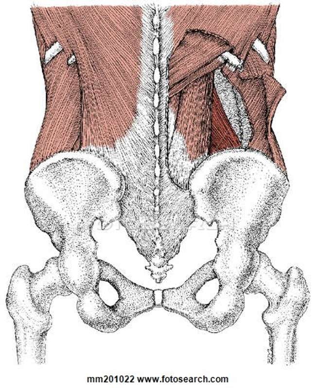
Illustration courtesy of - http://fscomps.fotosearch.com/online-courses/online-courses/bigcomps/LIF/LIF156/MM201022.jpg
Study Summary
| Study Design | Descriptive cross-sectional |
| Level of Evidence | III evidence from non-experimental descriptive studies, such as comparative studies, correlation studies and case-control studies |
| Participant Characteristics | Sample: Demographics
Inclusion Criteria:
Exclusion Criteria:
|
| Methodology | Trunk Muscle Strength Measurement
Measurement of lordosis and sacral angle
|
| Data Collection and Analysis |
|
| Outcome Measures |
|
| Results |
|
| Our Conclusions | The researchers' findings support the inclusion of E/F ratio assessment when working with individuals with chronic LBP. Further, the reduction in flexion strength relative to extension strength may be further evidence of the relative changes in activity described in the Lumbo Pelvic Hip Complex Dysfunction model (LPHCD). |
| Researchers' Conclusions | Lumbar lordosis is moderately correlated with trunk E/F strength ratio. This ratio can be applied as an indicator of strength imbalance during trunk muscle assessment. Though not the focus of the study, low E/F ratio was found to correlate with LBP. The researchers suggest that several other factors be examined before concluding that an abnormal lordotic angle can be directly associated with LBP. |
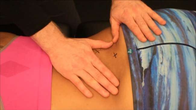
Erector spinae and multifidus static manual release by Dr. Brent Brookbush
Review & Commentary: This study adds to the body of research investigating correlations between anthropometric variables and chronic lower back pain (LBP). This research is unique in its use of computerized goniometer software to maximize inter-rater reliability of the lumbosacral angles. A relationship between trunk muscle strength ratios, lordotic and lumbosacral angles was observed in both men and women with LBP.
- The study had many methodological strengths:
- The researchers used validated methods - the B200 Isostation, a well-established means of measuring both isometric and isodynamic trunk strength (2), and the Cobb method, used to measure scoliosis curvature and was adapted in this study to measure lordotic angle.
- PACS software was used to measure the sacral angle, which increased inter-rater reliability over other studies that used manual measures.
- This study filled a gap in the body of research through its demonstration of a relationship between trunk muscle strength and spine angle in both men and women with LBP.
- Weaknesses that should be noted prior to clinical integration include:
- Participants were not selected randomly but rather chosen from a specific location after clinical evaluation, which limits the generalizability of the study. Drawing participants from the general population would provide a clearer comparison of variables in healthy and injured populations.
- This study had no intervention by design and was purely descriptive/observational. An intervention will be needed in future studies to build a stronger case for clinical integration.
- The B200 Isostation is not a device accessible to most human movement professionals, which limits the ability of clinicians and researchers to accurately replicate the study.
How This Study is Important:
The relationships between lumbosacral position, muscle strength and low back pain may highlight common compensatory patterns that influence assessment and intervention selection. This study compared changes in trunk muscle strength with lordotic curve and sacral slope angles, in those with low back pain and asymptomatic individuals. Prior studies have investigated lumbosacral position and muscle strength individually in those exhibiting signs low back pain; however, few studies have examined both in the same study (4-11). The inclusion of both outcome measures in this one study allows lumbosacral position and muscle strength to be correlated to one another. The correlation between low back pain, increased lumbar lordosis and sacral slope angle and a decrease in lumbar flexor endurance imply these issues should be considered collectively.
How the Findings Apply to Practice:
Human movement professionals should attempt to identify imbalances between trunk flexor and extensor strength, perhaps by identifying an “excessive lordotic curve ” during static and dynamic postures (example, Overhead Squat Assessment ) in those with low back pain. If an increased lordotic curve or muscle imbalance is identified, consideration should be given to rectifying this imbalance as part of a treatment/corrective program.
How does it relate to Brookbush Institute Content?
The findings of this study indicate that the ratio between trunk flexion and extension strength are correlated with chronic LBP. This is consistent with changes in muscle activity and interventions proposed in the Brookbush Institute's predictive models of Lumbo Pelvic Hip Complex Dysfunction (LPHCD) , Lumbosacral Dysfunction (LSD) , and Lower Extremity Dysfunction (LED) . Although this study did not employ an intervention, it establishes a relationship between E/F strength ratio and LBP. Human movement professionals should consider going beyond the generally recommended "core strengthening" to addressing the specific muscle weakness/movement impairments identified during assessment. Relative to this study, low back pain correlated with increased lumbar lordosis, may imply a need for greater focus on trunk flexor strength.
The following videos present a refresher on spine anatomy and trunk muscles, illustrate a common trunk flexor exercise and demonstrate a taping technique for low back decompression:
Muscles of the Core:
Quadruped Crawl
Plank:
Low Back Decompression Tape:
Bibliography:
- Andersson, G. B. (1999). Epidemiological features of chronic low-back pain . The lancet, 354(9178), 581-585.
- Gomez, T., Beach, G., Cooke, C., Hrudey, W., & Goyert, P. (1991). Normative database for trunk range of motion, strength, velocity, and endurance with the Isostation B-200 Lumbar Dynamometer. Spine, 16(1), 15-21. ABSTRACT
- van Middelkoop, M., Rubinstein, S. M., Kuijpers, T., Verhagen, A. P., Ostelo, R., Koes, B. W., & van Tulder, M. W. (2011). A systematic review on the effectiveness of physical and rehabilitation interventions for chronic non-specific low back pain . European Spine Journal, 20(1), 19.
- During, J., Goudfrooij, H., Keessen, W., Beeker, T. W., & Crowe, A. (1985). Toward Standards for Posture: Postural Characteristics of the Lower Back System in Normal and Pathologic Conditions. Spine, 10(1), 83-87.
- Evcik, D., & Yücel, A. (2003). Lumbar lordosis in acute and chronic low back pain patients. Rheumatology international, 23(4), 163-165.
- Christie, H. J., Kumar, S., & Warren, S. A. (1995). Postural aberrations in low back pain. Archives of physical medicine and rehabilitation, 76(3), 218-224.
- Hanson, D. S., Bridwell, K. H., Rhee, J. M., & Lenke, L. G. (2002). Correlation of pelvic incidence with low-and high-grade isthmic spondylolisthesis. Spine, 27(18), 2026-2029.
- Jackson, R. P., & McManus, A. C. (1994). Radiographic Analysis of Sagittal Plane Alignment and Balance in Standing Volunteers and Patients with Low Back Pain Matched for Age, Sex, and Size: A Prospective Controlled Clinical Study. Spine, 19(14), 1611-1618.
- Harrison, D. D., Cailliet, R., Janik, T. J., Troyanovich, S. J., Harrison, D. E., & Holland, C. (1998). Elliptical modeling of the sagittal lumbar lordosis and segmental rotation angles as a method to discriminate between normal and low back pain subjects. Clinical Spine Surgery, 11(5), 430-439.
- Marty C, Boisaubert B, Descamps H, Montigny JP, Hecquet J, Legaye J, Duval-Beaupère G.The sagittal anatomy of the sacrum among young adults, infants and spondylolisthesis patients.Eur Spine J2002 ; 11 : 119-125.
- Norton, B. J., Sahrmann, S. A., & Van Dillen, L. R. (2004). Differences in measurements of lumbar curvature related to gender and low back pain. Journal of Orthopaedic & Sports Physical Therapy, 34(9), 524-534.
© 2017 Brent Brookbush
Questions, comments, and criticisms are welcomed and encouraged -