
Research Review: Contributions of Lower Leg Muscles to Movement at the Ankle
By Jinny McGivern DPT, Certified Yoga Instructor
Edited by Brent Brookbush DPT, MS, PES, CES, CSCS, ACSM H/FS
Original Citation: Klein, P., Mattys, S., & Rooze, M. (1996). Moment arm length variations of selected muscles acting on talocrural and subtalar joints during movement: An in vitro study. Journal of biomechanics, 29(1), 21-30. ABSTRACT
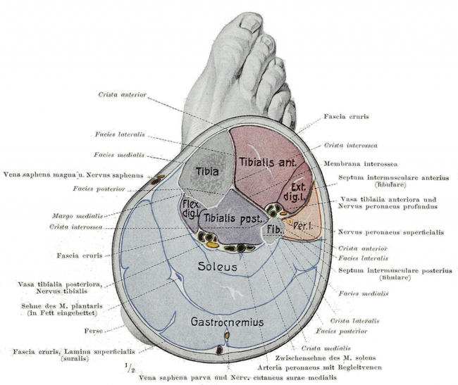
By Braus, Hermann - Anatomie des Menschen: ein Lehrbuch für Studierende und Ärzte, Public Domain, https://commons.wikimedia.org/w/index.php?curid=29934112
Why is this relevant?: This research study examined cadaver specimens in order to provide information about the contributions of specific muscles of the lower leg to joint actions at the ankle (talocrural and sub-talar joints). It also examined the relationship between those muscles with respect to the size of their moment arm and their ability to create movement at the ankle. To train optimal movement, it is essential to know the contribution of each muscle in a given movement pattern, whether the contribution of a muscle in a given pattern is optimal, and if not optimal, how that muscle is contributing to compensation.
Study Summary
| Study Design | Descriptive Study |
| Level of Evidence | Level VI: Evidence from a single descriptive or qualitative study |
| Subject Demographics |
|
| Outcome Measures | Morphometric data (from 9 specimens):
Moment arms of the following muscles in dorsiflexion/plantar flexion & inversion/eversion: |
| Results | Morphometric Data
Moment Arm Data At the talocrural joint:
At the sub-talar joint:
At the talocrural joint the following muscles demonstrated correlation between the size of their moment arms:
At the sub-talar joint the following muscles demonstrated correlation between the size of their moment arms:
There was not a significant relationship between talocrural joint position and moment arm of muscles at the sub-talar joint. |
| Conclusions | This research confirms that many of the muscles we traditionally consider prime movers for a given joint action are oriented to perform this role. Most of the muscles observed are each able to perform 1 motion per plane per joint; however, the TS demonstrated that it is the exception. During inversion at the sub-talar joint it demonstrated an eversion moment arm & in eversion an inversion moment arm. |
| Conclusions of the Researchers | Based on moment arm data, the TS appears to be the most important muscle in the performance of plantar flexion followed somewhat closely by FHL, and distantly by FL, FB and TP. FL & FB appear to function very similarly, and are most important for eversion. TP demonstrated the greatest ability to perform inversion, while TA was the only member of this group with a dorsiflexion moment arm. |
Image above indicates the location of the 2 joints observed in this study (note: that the label "True Ankle Joint" is given to the tibiotalor a.k.a talocrural joint - http://www.kidport.com/online-courses/online-courses/reflib/science/humanbody/skeletalsystem/Ankle.htm
Review & Commentary:
A strong point of this study was that 6 muscles were observed and compared to each other. This provides the human movement professional with a wealth of information about about the relationships between lower leg muscles and their contribution to movement. It answers questions such as, "Of all the muscles that are to perform a particular joint action, which has the greatest mass and mechanical advantage ?" This allows the human movement professional an opportunity to connect evidence and physics, with theoretical models of ankle motion. Another strong point of this article is the analysis by the authors of the pros & cons of various methods of quantifying the moment arms of individual muscles. The method that they ultimately selected allowed for changes in the axis of rotation as a movement was performed. This method matches well with functional human movement where the axis of joint rotation changes as a body part moves through a range of motion. The researchers utilized surgical pins to provide a sturdy fixation of 1 joint when observing moment arms of the other. While this provided useful information on isolated joint motion, isolated joint motion rarely occurs in human movement patterns. This aspect of the research methodology differed from function, but allows for the quantification of moment arms and force production at each joint. Analysis of the combined motion of the tibiotalor and sub-talar joints may be a good direction for future research. Additionally, the researchers did not comment on movement of, or lack of movement at the inferior tibio-fibular joint.
The greatest weakness of this research is that it was performed on cadaver subjects (in vitro), not on live subjects (in vivo). Further, the researchers utilized cadaver specimens (preparations of the lower leg and foot only) as opposed to a complete cadaver. We must use caution in how we apply the information gleaned from this data. The information is valuable and important, however it is essential to remember where it came from in the event that other authors report differing results in an in vivo situation. The authors state that previous research has noted little difference in arthrokinematics between in vivo and in vitro studies of the tarsal bones; however, it is uncertain how the behavior of soft tissue (especially fascia which was removed from the specimens) would have impacted the moment arms of the 6 tendons observed in this study.
This research highlighted a general difference in the behavior of muscles at the ankle. At the talocrural joint, as the ankle moves in the direction of the force vector the moment arm of the 6 muscles investigated increased. (e.g.. as dorsiflexion range increased, TA moment arm did as well). At the sub-talar joint, the inverse was true. (e.g. the moment arm of the muscles performing inversion decreased as the joint was moved into inversion) Speculation: From functional anatomy point of view, is it possible that greater strength is needed from muscles at the talocrural joint for the concentric phases of movement, whereas at the sub-talar joint greater strength is needed during eccentric phases of moment, thus the greater moment arm at earlier points in the range? Further research is needed to further understand this relationship. Further research is also needed to determine moment arms for Extensor Hallucis Longus, Extensor Digitorum Longus , Flexor Digitorum Longus.
Why is this study important?
This study provides information on which muscles (of the ones studied) are optimally positioned to perform a joint action in the sagittal and/or frontal plane at the talocrural & sub-talar joints. As our understanding of muscle behavior improves, this dictates both how we assess human movement and how we approach our intervention plans.
How does it affect practice?
Practically, this information assists us in understanding which muscles are considered the prime movers for a given joint action, and also points out which muscles may potentially become "overactive synergists " when the prime mover is not recruiting optimally. For example, the data indicates that TP has the largest moment arm for inversion. In the event that TP is not recruiting optimally for a given task, FHL may take on the role of overactive synergist because it is set up to have the second best mechanical advantage to perform inversion of the muscles observed in this research.
In addition to identifying prime movers this research also identifies which muscles oppose a given joint action, the antagonists. By virtue of studying several muscles, the authors provide us with a big picture understanding of the balance of forces around a given joint. For example, of the muscles studied, only the TA demonstrated a dorsiflexion moment arm; however, 5 muscles were observed to exhibit moment arms for plantar flexion. Even if we include the 2 other muscles which are believed to assist in dorsiflexion (extensor hallucis longus and extensor digitorum longus ), there remains an "imbalance" in the setup of the muscular system around the talocrural joint. This may explain why we often observe restrictions in dorsiflexion ROM, but rarely see limitations in plantar flexion ROM.
How does it relate to Brookbush Institute Content?
This research supports the model of Lower Leg Dysfunction (LLD) described by the Brookbush Institute. It describes how the mechanical advantage (described by moment arms) of different muscles around the ankle joint may lead to tendencies for plantar flexors and evertors to become short and overactive, whereas the dorsiflexors and invertors tend to become long and under-active. The ability of the TS to perform both eversion and inversion helps to explain an incongruency in the LLD model regarding over-activity of the soleus despite this muscle traditionally being thought of as an invertor.
As discussed above understanding which muscles are considered the prime movers for a given joint action, may also help indicate which muscles become "overactive synergists " (long and overactive) when the prime mover is not recruiting optimally. Once dysfunction is identified, the next question of the human movement professional becomes "What do I do about it?"
The Brookbush Institute provides suggestions for intervention designed to correct Lower Leg Postural Dysfunction. To continue the example above with TP as an inhibited prime mover & FHL as an overactive synergist, it is necessary to design an exercise that activates the TP while inhibiting the FHL . The Brookbush Institute approaches this conundrum by suggesting TP activation exercise where the individual performs heel raise with inversion, while also extending the toes (Video below). Toe extension reciprocally inhibits the FHL by increasing activity of the EHL , where it does not impact TP in any way because TP does not cross the metatarsal joints. Ideally, this decreases synergist activity, and increases prime mover motor unit recruitment resulting in more optimal movement pattern.
Fibularis Longus and Brevis SA Active Release
Calf and Fibularis (Peroneal) SA Static Stretching Technique
Dynamic Calf Stretch - Gastrocnemius, Soleus & Fibularis muscles (Peroneals)
Ankle Mobilization
Tibialis Anterior Isolated Activation
Posterior Tibialis Isolated Activation
Posterior Tibialis Activation Progression
Tibialis Anterior Reactive Integration Progressions
© 2014 Brent Brookbush
Questions, comments, and criticisms are welcomed and encouraged -