
Research Review: The Role of Tibialis Posterior Fatigue on Foot Kinematics During Walking
By David Chessen DPT, PT, OCS, MBA
Edited by Brent Brookbush DPT, PT, COMT, MS, PES, CES, CSCS, ACSM H/FS
Original Citation: Pohl, M. B., Rabbito, M., and Ferber, R. (2010). The role of tibialis posterior fatigue on foot kinematics during walking. Journal of Foot and Ankle Research, 3(1), 6. ABSTRACT
Why the Study Is Relevant: The tibialis posterior serves as an invertor of the rear foot and provides dynamic arch support for the mid foot (1). The effect of tibialis posterior dysfunction on the mechanics of ambulation has been well documented (2,3). However, the effect of tibialis posterior fatigue on foot kinematics in healthy individuals has received little attention. This 2010 study investigated the effect of fatiguing the tibialis posterior through resisted foot adduction on foot kinematics during walking.
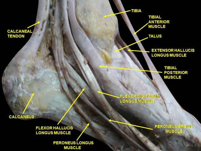
Tibialis Posterior Tendon Source: https://brentbrookbush.com/articles/anatomy-articles/muscular-anatomy/tibialis-posterior/
Study Summary
| Study Design | Single-group quasi experimental |
| Level of Evidence | IIB evidence from at least one other type of quasi-experimental study |
| Participant Characteristics | Demographics
Inclusion Criteria:
Exclusion Criteria:
|
| Methodology |
|
| Data Collection and Analysis |
|
| Outcome Measures | Kinematic variables of interest:
|
| Results | Force output:
Kinematics:
Structure and Kinematics:
|
| Our Conclusions | This study found no significant changes in rear foot eversion during walking following a fatiguing exercise protocol for the tibialis posterior. This study has influenced the Brookbush Institute (BI) model for lower extremity dysfunction (LED); resulting in consideration of labeling pronation as the osteokinematic change correlated with "feet flatten" rather than eversion. |
| Researchers' Conclusions | An exercise protocol aimed at fatiguing the tibialis posterior produced no substantial changes in rear foot eversion during walking. No significant changes in forefoot kinematics were observed after the fatiguing protocol. (Note: medial longitudinal arch height was not measured) |
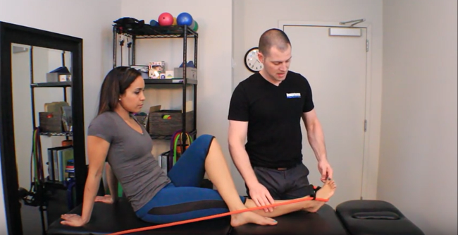
Tibialis Posterior Activation
Tibialis posterior activation exercise from the Brookbush Institute. Source: https://brentbrookbush.com/articles/corrective-exercise-articles/activation/tibialis-posterior-activation/
Review & Commentary:
This study found that a fatiguing exercise protocol for the tibialis posterior did not alter peak rear foot and forefoot kinematics with walking; however, greater excursion of the rear foot was noted.
The study had many methodological strengths, including:
- The study used a clinically relevant exercise in foot adduction to fatigue the tibialis posterior .
- The timeline for the fatiguing protocol was well documented and explained allowing future replication of the study methods.
- To our knowledge this is the first study to measure the effect of a fatiguing protocol for the tibialis posterior on gate.
Weaknesses that should be noted prior to clinical integration of the findings include:
- No electromyographic (EMG) analysis was performed making it difficult to determine whether the tibialis posterior was directly affected by the fatiguing exercise protocol (and what effect fatigue has on activity).
- The contribution of other structures or muscles was not analyzed which may have affected the results of this study.
- Structural measures of the participants' medial longitudinal arch and forefoot were not measured.
How This Study is Important:
The findings of this study suggest that other muscles may support the rear foot, and/or that the tibialis posterior does not have a significant influence over rear foot mechanics. Further studies should consider EMG of synergistic muscles and inclusion of the navicular drop test to assess forefoot mechanics.
How the Findings Apply to Practice:
Rear foot mechanics (eversion) may not be a reliable assessment of tibialis posterior activity and strength. Human movement professionals should assess (and intervene when appropriate) the entire ankle/foot complex and consider all potential structures contributing to the altered mechanics notes.
How does it relate to Brookbush Institute Content?
The tibialis posterior is noted as being long and under active in the Brookbush Institute (BI) predictive model of lower extremity dysfunction (LED) . Activation techniques are suggested by the BI to enhance the functioning of the tibialis posterior and improve movement dysfunctions such as "feet flatten ." The study did not find significant changes in foot mechanics or rear foot eversion with fatiguing of the tibialis posterior , which suggests that other structures may support and compensate for the fatigued tibialis posterior . The tibialis anterior also provides muscular support for the medial longitudinal arch, acting as a powerful supinator of the foot (4), which may imply that "feet flatten " is the result of forefoot pronation and not rear foot eversion. It is important to note that this study investigated asymptomatic individuals with no history of LED . It is unknown whether similar results would have occurred in symptomatic individuals.
The following videos illustrate common assessment techniques and interventions used to affect the tibialis posterior :
Tibialis Posterior Manual Muscle Testing
Tibialis Posterior Activation
Tibialis Posterior Activation Progression 2
Bibliography:
- Tome, J., Nawoczenski, D. A., Flemister, A., & Houck, J. (2006). Comparison of foot kinematics between subjects with posterior tibialis tendon dysfunction and healthy controls. Journal of Orthopaedic & Sports Physical Therapy, 36(9), 635-644.
- Ness, M. E., Long, J., Marks, R., & Harris, G. (2008). Foot and ankle kinematics in patients with posterior tibial tendon dysfunction. Gait & posture, 27(2), 331-339.
- Ringleb, S. I., Kavros, S. J., Kotajarvi, B. R., Hansen, D. K., Kitaoka, H. B., & Kaufman, K. R. (2007). Changes in gait associated with acute stage II posterior tibial tendon dysfunction. Gait & posture, 25(4), 555-564.
- Klein, P., Mattys, S., & Rooze, M. (1996). Moment arm length variations of selected muscles acting on talocrural and subtalar joints during movement: An in vitro study. Journal of biomechanics, 29(1), 21-30.
© 2017 Brent Brookbush
Questions, comments, and criticisms are welcomed and encouraged -