
Research Review: Gluteal Muscle Activation During Common Rehabilitation and Injury Prevention Exercises
By Tristan Rodik, M.AT., ATC
Edited By Brent Brookbush, DPT, PT, COMT, MS, PES, CES, CSCS, ACSM H/FS
Original Citation: DiStefano, L. J., Blackburn, J. T., Marshall, S. W. and Padua, D. A. (2009) Gluteal muscle activation during common therapeutic exercises. Journal of Orthopaedic and Sports Physical Therapy, 39(7), 532-540. ABSTRACT
Why the Study is Relevant: Underactivity of the gluteus maximus and gluteus medius (glute complex ) has been correlated with low-back pain and lower extremity dysfunction , including patellofemoral pain syndrome and ankle sprains (1-8). This 2009 study used surface electromyography to investigate the muscle activation patterns of 12 rehabilitation exercises commonly selected by human movement professionals to target the glute complex . Human movement professionals may the findings of this study to aid in the optimal selection and progression of glute complex activation exercises.
Study Summary
| Study Design | Cross-sectional Study |
| Level of Evidence | IIA Evidence from at least one controlled study without randomization |
| Subject Characteristics | Demographics
Inclusion Criteria:
Exclusion Criteria:
|
| Methodology |
|
| Data Collection and Analysis |
|
| Outcome Measures |
|
| Results |
|
| Our Conclusions | The results support including clam shells and side-lying hip abduction exercises into gluteus medius isolated activation protocols. The findings also suggest that the gluteus maximus is more active in weight-bearing exercises, likely due to increased load. |
| Researchers' Conclusions
|
The side-lying hip abduction exercises represented the highest %MVIC for the gluteus medius. The single-limb squat and single-limb deadlift exercises represented the highest %MVIC for the gluteus maximus. Performing these exercises may improve rehabilitation efficiency and prevention programs. |
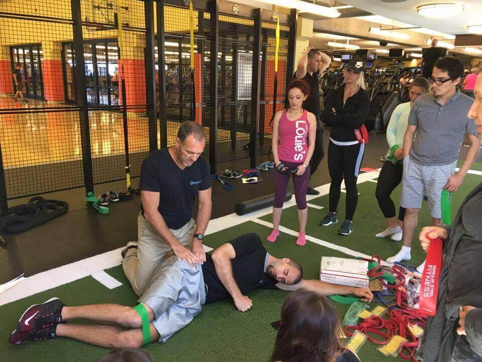
Dr. Brent Brookbush demonstrating positioning to manual muscle test the gluteus medius (Image: Courtesy of the Brookbush Institute ©)
Review and Commentary
This study adds to a growing body of research comparing gluteus medius and gluteus maximus exercises using electromyography (EMG) (10, 11). The greatest percentage of gluteus medius maximal voluntary isometric contraction (%MVIC) was noted during the side-lying hip abduction exercise. The greatest %MVIC for gluteus maximus was the single-leg deadlift and single-leg squat. It is hypothesized that these exercises resulted in the greatest %MVIC due to greater lever length/increased load and increased stability demand.
This study had many methodological strengths, including:
- Intra-class coefficients (ICC) demonstrated moderate to high reliability across trials of the 12 exercises. This confirms that the electrodes for electromyographic analysis were accurately placed on all participants.
- Exercises were performed in random order, reducing the potential for a learning effect due to similarities among some exercises. The random order also reduced the risk of fatigue influencing findings.
- The exercises evaluated in this study are commonly used by human movement professionals, increasing applicability.
Weaknesses that should be noted prior to clinical integration:
- All participants reported no pain or surgery within the past two years. The %MVIC may differ for injured or post-surgical clients.
- MVIC testing was conducted after participants completed all 12 exercises, this may have resulted in fatigue and an increase in recorded %MVIC.
- Aside from lateral band walks, external resistance was not added for the exercises. It is unclear whether adding weight would further increase %MVIC, and/or modify the order of exercise based on %MVIC.
Why This Study is Important:
The importance of gluteus medius and gluteus maximus strengthening and its correlation to lower-extremity function and pain has been well-researched (1-8, 9, 12). This comparative, practical study provides objective EMG data on the differences between various commonly used exercises for gluteus medius and gluteus maximus strengthening. More research of this type is desperately needed for all exercises, techniques and modalities. The following exercises were tested:
- Side-Lying Hip Abduction
- Single-Limb Squat
- Single-Limb Deadlift
- Lateral Band Walks
- Multiplanar Lunges (forward, sideways and transverse )
- Multiplanar Hops (forward , sideways and transverse )
- Clamshells at 30° and 60°
How the Findings Apply to Practice:
The findings of this study demonstrate which exercises result in the greatest %MVIC for gluteus maximus and gluteus medius activation and several exercises that surpass the recommended 50-60 %MVIC for strengthening (13). The side-lying hip abduction exercise for gluteus medius , single-leg deadlift , and single-leg squat for gluteus maximus achieved the highest %MVIC. Lateral band walks and sideways hop for the gluteus medius , and the transverse plane lunge for the gluteus maximus also surpassed the 50-60 %MVIC threshold. Human movement professionals should consider integrating these exercises into any program with the intent of increasing glute complex strength, and further consider lever length/load and stability demands in the progression of exercise. Exercises listed in order of %MVIC below:
- The following exercises scored the highest for gluteus medius (%MVIC +/- Standard Deviation (SD)):
- Side-lying hip abduction (81 +/- 42)
- Single-limb squat (64 +/- 24)
- Lateral band walk (61 +/- 34)
- Single-limb deadlift (58 +/- 25)
- Sideways hop (57 +/- 35)
- The following exercises scored the highest for gluteus maximus (%MVIC +/- SD):
- Single-leg squat (59 +/- 27)
- Single-limb deadlift (59 +/- 28)
- Transverse plane lunge (49 +/- 20)
- Forward lunge (44 +/- 23)
- Sideways lunge (41 +/- 20)
How this Study Relates to Brookbush Institute Content:
The Brookbush Institute (BI) has used studies like this one to refine progressions for gluteus medius activation and gluteus maximus activation . Weakness/inhibition of these muscles is noted in the predictive models of Lower Extremity Dysfunction (LED) , Lumbo-pelvic Hip Complex Dysfunction (LPHCD) , and Sacroiliac Joint Dysfunction (SIJD) . It is BI's assertion that further improvements in muscle recruitment and motion may be achieved by adding release and lengthening techniques , as well as joint mobilizations (when appropriate) prior to isolated activation techniques. Once activation has been achieved, clients may progress to subsystem integration for the posterior oblique subsystem . The following assessments and exercises are recommended to identify and address gluteal complex underactivity.
Functional Anatomy of the Gluteus Maximus, Medius and Minimus
Manual Muscle Testing for the Gluteus Medius
Manual Muscle Testing for the Gluteus Maximus
Glute Activation Circuit:
Gluteus Medius Activation Progressions
Side Stepping Progressions for Gluteus Medius Reactive Activation
Gluteus Maximus Reactive Activation
Recommended Readings:
- Electromyographic data of common exercises for the gluteus medius and gluteus maximus .
- Electromyographic data and biomechanical considerations for exercise progressions of the gluteus medius .
Bibliography:
- Bolgla, L. A., Malone, T. R., Umberger, B. R. and Uhl, T. L. (2011) Comparison of hip and knee strength and neuromuscular activity in subjects with and without patellofemoral pain syndrome. The International Journal of Sports Physical Therapy, 6(4), 285-296.
- Bolgla, L. A., Malone, T. R., Umberger, B. R. and Uhl, T. L. (2008) Hip strength and hip and knee kinematics during stair descent in females with and without patellofemoral pain syndrome. Journal of Orthopaedic and Sports Physical Therapy, 38(1), 12-18
- Souza, R. B. and Powers, C. M. (2009) An evaluation of hip strength and femoral structure in women with and without patellofemoral pain. The American Journal of Sports Medicine, 37(3), 579-587. doi: 10.1177/0363546508326711
- Ireland, M. L., Wilson, J. D., Ballantyne, B. T. and Davis, I. M. (2003) Hip strength in females with and without patellofemoral pain. Journal of Orthopaedics and Sports Physical Therapy, 33, 671-676
- Ramskov, D., Barton, C., Nielson, R. O. and Rasmussen, S. (2015) High eccentric hip abduction strength reduces the risk of developing patellofemoral pain among novice runners initiating a self-structured running program: a 1-year observational study. Journal of Orthopaedic and Sports Physical Therapy, 45(3), 153-161
- Yoo, W. G. (2014) Effect of the individual strengthening exercises for posterior pelvic tilt muscles on back pain, pelvic angle, and lumbar ROM of a LBP patient with excessive lordosis: a case study. Journal of Physical Therapy Science, 26(2), 319-320
- Leinonen, V., Kankaanpaa, M., Airaksinen, O. and Hannine, O. (2000) Back and hip extensor activities during trunk flexion/extension: effects of low back pain and rehabilitation. Archives of Physical Medicine and Rehabilitation, 81(1), 32-37
- Bullock-Saxton, J. E. (1994) Local sensation changes and altered hip muscle function following severe ankle sprain. Physical Therapy, 74(1), 17-28
- Dolak, K. L., Silkman, C., Medina McKeon, J., Hosey, R. G., Latterman, C. and Uhl, T. L. (2011) Hip strengthening prior to functional exercises reduces pain sooner than quadricep strengthening in females with patellofemoral pain syndrome: a randomized clinical trial. Journal of Orthopaedic and Sports Physical Therapy, 41(8), 560-570
- Boren, K., Conrey, C., Le Coguic J., Paprocki, L., Voight, M. and Robinson K. (2011) Electromyographic analysis of the gluteus medius and gluteus maximus during rehabilitation exercises. International Journal of Sports Physical Therapy, 6(3), 206-223
- Bolgla, L. A. and Uhl, T. L. (2005) Electromyographic analysis of hip rehabilitation exercises in a group of healthy subjects. Journal of Orthopaedic and Sports Physical Therapy, 35(8), 487-494
- Cooper, N., Scavo, K., Strickland, K., Tipayamongkol, N., Nicholson, J., Bewyer, D., Sluka, K. (2015) Prevalence of gluteus medius weakness in people with chronic low back pain compared to healthy controls. European Spine Journal, 25(4), 1258-1265
- Atha, J. (1981) Strengthening muscle. Exercises and Sports Sciences Reviews, 9, 1-73
- Ayotte, N. W., Stetts, D. M., Keenan, G. and Greenway, E. H. (2007) Electromyographical analysis of selected lower extremity muscles during 5 unilateral weight-bearing exercises. Journal of Orthopaedics and Sports Physical Therapy, 37(2), 48-55
- Myers, J. B., Pasquale, M. R., Laudner, K. G., Sell, T. C., Bradley, J. P. and Lephart, S. M. (2005) On-the-field resistance-tubing exercises for throwers: an electromyographic analysis. Journal of Athletic Training, 40(1), 15-22
© 2017 Brent Brookbush
Questions, comments and criticisms are welcomed and encouraged.